The OBC has a FULL license to Imaris 3/4D Image Visualization and Analysis Software on a high end workstation, directly linked to the OBC server for convenient data access for analysis.
Imaris has a free version, Imaris Viewer, that is a free 3D/4D microscopy image viewer for viewing raw images as well as those analysed within Imaris.
- Browse files as thumbnails and open 3D images
- Visualize datasets previously analyzed in Imaris
- Interact with 3D images using intuitive mouse controls
- Inspect your images with clipping planes and 2D slicers
- Easily generate high-resolution snapshots
- Adjust and save brightness and contrast
Imaris Full comes with Visualization of complex 3/4D microscopy datasets with automated Spots and Surfaces detection and visualisation (100s of GBs), smart detection of complex objects, tracing of neurons, blood vessels (no lumen) or other filamentous structures, tracking including cell division detection, batch analysis and a wide range of customized analysis powered by MatLab or Python.
For tutorials on how to use the different Imaris modules, go to Imaris Learning.
During the COVID-19 pandemic Imaris has held some very helpful webinars. below are links to the talks
“Importing Data Into Imaris And The Basics Of Image Visualization” here.
“All you need to know about 3D Rendering and Visualization in Imaris” here.
“Create Eye-Catching Animations and Snapshots using Imaris” here.
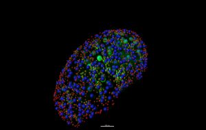
Pink: Oocytes of primordial follicles (<12 um)
Yellow: Oocytes of primary follicles (<25 um)
Blue: Oocytes of secondary follicles (<65 um). Image courtesy of Dr. Jinhwan Lim in Professor Ulrike Luderer’s Lab