The Optical Biology Core Facility would like to announce that the Light sheet fluorescence microscopy (LSFM) system is now available to all trained users
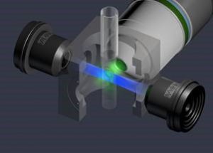
The Zeiss Z1 Light sheet microscope uses 2 objective lenses perpendicular to each other to excite/detect the sample. The illumination optics create a single sheet of light through a plane of the sample, which is then detected with a s-CMOS camera PCO.edge, max frame rate 30 fps at 960 x 1024 pixel resolution, pixel size 6.5 μm, maximal 3.8 MPixel at lower frame rates. This allows for rapid imaging of thick tissue samples, with very little bleaching and photo-toxicity. The Z1 light-sheet can image both live samples (in aqueous media,) and cleared tissues (in 1.33/1.45/1.55 refractive index media), and has 4 laser lines for excitation (405nm, 488nm, 561nm, and 633nm) and 2 cameras. Additional details can be found HERE
Training for new users began 05/21/2018. Users should bring samples and at least 25ml of buffer to fill the imaging chamber.
Before coming in for training, all users should watch the tutorial videos on the Zeiss Knowledge base .
So far, we have had great success doing time lapse imaging on zebrafish, and cleared tissues (brain, heart, mammary tissue, skin, ovaries, spinal chord) cleared with many different clearing techniques, including iDisco+, Cubic, Scale, Clarity etc. Whole mouse brain imaging is possible thanks to a custom 2.5x imaging objective and large chamber made by Translucence Bio (part of a UC Irvine start up company).
For more examples on how to mount samples for the Z1, check out the attached white paper from Zeiss. Lightsheet-Z.1_sample-preparation
Funding for the microscope was provided by the Vice Chancellor for Research, the Charlie Dunlop School of Biological Sciences, the Samueli School of Medicine, the Departments of Developmental & Cell Biology, Neurobiology & Behavior and Pharmaceutical Sciences, the Center for the Neurobiology of Learning and Memory and the Chao Comprehensive Cancer Center.
For more information or to schedule a time for training contact Dr Adeela Syed (adeelas@uci.edu)