The OBC provides access to state-of-the-art light microscopes (confocal, mesoscale lightsheet, and Super Resolution), as well as software for 3D and 4D analysis. All instruments are accessible 24/7 to trained researchers of laboratories across the campus and the local research community. All researchers are trained by Dr. Adeela Syed with additional imaging support provided as needed
The OBC now uses ilabs for scheduling and updating researchers on the OBC latest news!
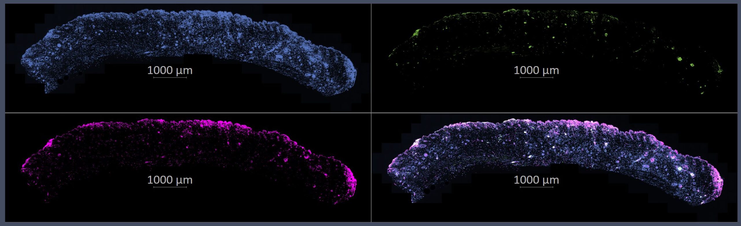
July 2025: NEW Zeiss Axioscan Z.1 slide scanner is here and ready for use! The image below was taken with 40x Air objective, over 200 tiles, in about 15 minutes! Skin section with DAPI, the 488 is CD45 which marks immune cells, and the 647 is y-H2AX, which marks cells with DNA damage.
Image courtesy of Kiarash Forouzesh in Dr. Bogi Andersen’s lab
The Optical Biology Core Facility (OBC), founded in 1994, is directed by Dr. Rahul Warrior, Professor, Department of Developmental & Cell Biology, and is managed by Dr. Adeela Syed (Optical Biology Core), Dr. Michelle Digman (Laboratory of Fluorescence Dynamics) Dr. Mihaela Balu (Non Linear Optical Microscopy NLOM) and Dr. Michael Hou (Flow Cytometry Facility)
1. A self use facility located in 4443 McGaugh Hall at the UC Irvine Campus houses a total of 7 microscopes (4 confocals, 1 Mesoscale Lightsheet, 1 Super Resolution, 1 Slide Scanner) and 5 workstations:
OBC Self-Use Facility
Located in McGaugh Hall at UC Irvine, the OBC Self-Use Facility provides 24/7 access to a suite of high-performance commercial and custom-built imaging systems. Trained users can perform both fixed and live imaging across a wide range of advanced microscopy modalities and quantitative analysis workflows.
Available instrumentation includes:
Zeiss LSM 980: Equipped with a two-photon laser, 32-channel spectral detector, dual NDDs, and high-sensitivity Airyscan for deep tissue and high-speed imaging. Includes an environmental chamber for live imaging.
Zeiss LSM 900 Airyscan 2: Offers fast, high-resolution imaging with reduced phototoxicity. Also includes an environmental chamber for live-cell imaging.
Leica SP8: Features six laser lines (405, 458, 488, 514, 561, 633 nm), two HyD and two PMT detectors, and 20x multi-immersion and 63x oil objectives.
Zeiss LSM 780: Includes a two-photon laser, six standard laser lines (405–633 nm), and a 32 channel spectral detector.
Zeiss Elyra 7 Lattice SIM: Provides 3D SIM imaging with SIM² processing and single-molecule localization (SMLM) capabilities. Includes an environmental chamber for live imaging.
Zeiss Lightsheet Z1: Enables live and cleared tissue imaging with four laser lines (405, 488, 561, 633 nm) and three chambers, including one optimized for large, optically cleared samples in high refractive index media. To get started in tissue clearing and staining methods check out kits and services from our local Translucence Bio
Zeiss Automated Slide Scanner: Supports high-resolution, multi-channel IHC/IF imaging (up to 7 colors) with AI Tissue Finder and ZEN Connect integration for seamless workflow across systems.
Available image analysis software:
Imaris 3/4D (full license) – for 3D/4D visualization and quantification
Arivis Vision4D – for large-volume image analysis
ZEN Black for SIM processing, – for SIM acquisition and processing
ZEN Blue for Airyscan processing – for Airyscan processing
Stitchy – for Lightsheet image stitching and rendering
Supported imaging capabilities:
Confocal microscopy
Two-photon microscopy
Super-resolution (Lattice SIM with SIM2 processing and SMLM: dSTORM, DNA-PAINT)
Light sheet mesoscale imaging
Comprehensive one-on-one training and regularly hosted workshops prepare users to take full advantage of the facility. Trained users may reserve and access instruments 24 hours a day, 7 days a week.
More information on scheduling and recharges can be found on the iLabs scheduling site. Users can sign up for time on the scopes 24hours/day, 7 days a week.
2. The Nonlinear Optical Microscopy (NLOM) Lab located in the Beckman Laser Institute and Medical Clinic
This facility is equipped with state-of-the-art commercial and in-house optical microscopy platforms for biological and biomedical imaging. The imaging platforms provided as shared resources in this facility are:
- Leica SP8 Falcon + coherent anti-Stokes Raman Scattering (CARS) for confocal and nonlinear optical microscopy. This is a customized commercial imaging platform that includes the following modalities: confocal and two-photon excited fluorescence (TPEF), second harmonic generation (SHG), CARS and fluorescence lifetime microscopy.
The NLOM Lab provides collaborative opportunities in technology development to improve multiphoton microscopy (MPM) based imaging with large fields of view and rapid scanning for monitoring skin therapies, non-invasive diagnosis of skin cancers and other skin conditions.
Dr. Mihaela Balu directs the NLOM Lab and provides consultation and protocol development for investigators seeking new imaging applications.
Users are supported by extensive shop facilities that allow construction and modification of imaging platforms (https://sites.uci.edu/nlom/).
3. The Flow Cytometry Facility (FCF) is located in 3101 Hewitt hall.
The Institute for Immunology now offers latest in flow cytometry services at our Flow Core Facility. The facility provides the latest technology and professional technical assistance for flow cytometric analysis and sorting. Our suite of multi-parameter flow cytometers are well equipped for fluorescence-activated cell sorting (FACS) and emerging flow cytometry assays. Dr. Michael Hou manages the facility and is available for support and training. https://sites.uci.edu/ififlowcore/